 EN
EN  DE
DE  ES
ES  FR
FR 
Diagnosis of Functional Hallux Limitus can be made upon clinical examination using a specific test called the "Flexor Hallucis Longus (FHL) stretch test".
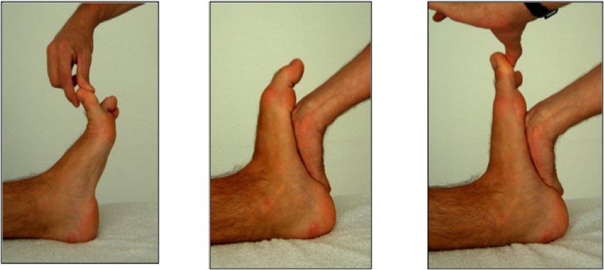
The FHL stretch test is carried out in 3 phases:
- Phase 1 : Patient lying on their back, ankle in plantar flexion position, free mobility of the MTP1 joint is ensured
- Phase 2 : The ankle is placed in maximum dorsiflexion by a firm support under the first metatarsal rays, (using palm of the hand and support with the weight of the body).
- Phase 3 : Exert a dorsiflexion on the MTP joint of the 1st ray by pushing the toe back.
If you can push the toe back, the test is negative. If, on the other hand, as in the image above, you are unable to push the toe back, the test is positive. A positive test shows the blockage of the tendon sliding of the FHL in the retro-talar tunnel at the level of the hindfoot.
As a corollary to this test, we have shown that it’s possible to recover the FLH tendon sliding by mobilizing the subtalar joint with the so-called “vacuum cord” maneuver (analogy of a blocked cord at an angle, or under a table leg which must first be released in order to slide again) (ref). The maneuver consists of unblocking the talocalcaneal joint by exerting traction in the axis of the leg, accompanied by small shearing movements.
A click is felt, synonymous with joint unlocking, and the space created by the joint distraction gives the tendon the possibility of sliding again. This sliding is perceived by the patient during the stretch test in the form of a tension in the calf along the muscle of the FHL or sometimes in the retro-malleolar region at the muscular-tendon junction. This maneuver associated with the stretching exercises of the FHL constitutes the basis of the orthopedic treatment.
Other clinical signs very suggestive of FLH:
- Tendon of the FHL is painful on palpation under the sole of the foot
- Hyperkeratosis on the inner side of the pulp of the big toe.
- Compensatory hyperextension of the interphalangeal gland.
- Absence of keratosis under the head of the first metatarsal.
- Marked keratosis under the head of the 5th metatarsal.
- A claw of the 2nd toe.
- Blockage of the subtalar joint.
- Wear of the shoe on the outer side of the heel.
- Wear of the sock or the insole under the pulp of the big toe.