General points
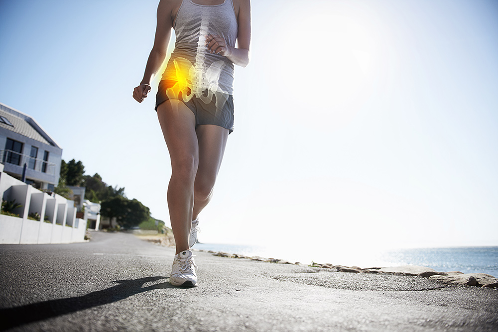
To understand the implication of the FHL on the hips, you have to study movement. When walking, the knee functions as a hinge joint just like the ankle. The rotational component is given by the hip upstream and by the subtalar downstream. These two joints by their anatomical common points are called "ball-in-the-socket joints" in English, “coxae pedis” and “femoris” by Professor Pisani who has wonderfully explained the implication of the coxa pedis in the mechanics of walking. The two joints are interdependent. If one of them is blocked, the other must follow the imposed direction of movement or try to resist it by the force of the stabilizing muscles. This is precisely what happens in the FHL where the subtalar is blocked. The hip is trained in internal rotation (medial collapse) during the support phase and in external rotation in the taligrade phase with a gluteal lever arm which loses its effectiveness.
The hip and how to assess its mobility usually involves measuring the movement of the femur in relation to the pelvis. However, to have a dynamic view of the hip, it must be looked upon as the movement of the pelvis over a single femoral head because that is how we function when walking. The advantage of using this reference (which is not natural because it is not what our eyes see) is that we can better appreciate and visualize the muscle vectors during movement, in particular for the gluteus medius. This muscle is crucial in stabilizing the pelvis and its lever arm is modified in the context of the FHL.
The dysfunction induced by FHL is global and involves the lumbopelvic statics where the blockage is reflected in the sagittal plane by an increased bending moment. The pelvis tilt forward is almost constant unless there is sufficient muscle gain to counter this tendency. In the rotary plane, the shift of the moments of transition in pro-supination is reflected by a medial collapse of the hip in adduction-internal rotation during the support phase and an exaggerated external rotation of the femur during the attack of the step. The consequences of this dynamic imbalance lead to overload lesions that affect the cartilage or the labrum (fibrocartilaginous bead that surrounds the hip) and which can lead to osteoarthritis.
Learn more
Consequences of hip dysfunction and differential diagnosisGluteal insertion tendinopathies or lesions of the hip cuff
 EN
EN  DE
DE  ES
ES  FR
FR 